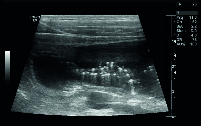
Ultrasound (sonography)
Four modern ultrasound devices with high resolution are available to our patients. Among other things they also enable Doppler examinations for the assessment of blood vessels. The images from the examination are saved digitally and can be compared during control examinations directly with current examination results. The ultrasound examination of the abdominal cavity enables assessment of the internal structure of such organs as, for example, the liver, spleen, gastrointestinal tract as well as urinary organs and genitals. After a short preparation of the patient, which includes the shearing of the abdomen and the application of the contact gel, the ultrasound examination can be performed mostly without anesthesia. In many cases a diagnosis can be determined at the same time.
In case of other diseases, the ultrasound however enables a targeted, minimally invasive cell or tissue sampling from changed organs. As such, structures that have already changed by just several millimeters can be examined and a diagnosis can be determined by means of microscopic evaluation. Since some diseases in the initial stage can only be detected using ultrasound, this imaging technique is an element of an extended medical check-up. Therefore, ultrasound is a perfect addition to X-ray diagnostics and blood test. In case of specific changes in the thoracic cavity that are determined using X-ray, the ultrasound enables a further restriction of the disease process, as well as a targeted sample collection, which can lead to a definite diagnosis.
For the diagnostics of heart diseases, an ultrasound examination is also required in most cases.
To cardiology


